Using Ultrasound to Heal: Therapeutic Ultrasound and Musculoskeletal Regeneration
There are two general types of medical ultrasound: diagnostic and therapeutic. Diagnostic ultrasound is widely used to produce real-time images of the body, including for the determination of musculoskeletal injuries and follow-up of musculoskeletal interventions. Contrastingly, therapeutic ultrasound produces bioeffects within the body, via the direct or indirect interaction of acoustic waves with cells/tissue, with the goal of benefiting patient health. Ultrasound-generated bioeffects are caused by mechanical and/or thermal phenomena such as acoustic streaming, cavitation, and heating. The ability of ultrasound to produce bioeffects is highly dependent on the characteristics of the ultrasound wave (e.g., frequency, pressure, duty cycle), the anatomical location of exposure, and tissue pathology. The first publication on ultrasound-generated bioeffects was in 1927 [1]. Since then, the field of therapeutic ultrasound has grown immensely, revolutionizing the treatment of a myriad of diseases including cancer, essential tremor, uterine fibroids, Parkinson’s disease, and kidney stones. Like diagnostic ultrasound, therapeutic ultrasound is non-invasive. In many applications, the ultrasound used for therapy is highly focused, thereby enabling the generation of bioeffects with millimeter or sub-millimeter spatial resolution.
Low-intensity pulsed ultrasound (LIPUS) is an example of therapeutic ultrasound clinically approved for use as a musculoskeletal intervention, specifically with radius and tibial fractures as well as non-unions. LIPUS can accelerate bone healing based on the stimulation of mechanotransductive pathways (e.g., MAPK, integrins), though the exact mechanisms involved are not fully understood [2].
There are many exciting preclinical applications of therapeutic ultrasound within musculoskeletal research. Therapeutic ultrasound has been used to target administered mesenchymal stromal cells (MSCs) to muscle based on the local enhancement of cytokines and growth factors [3,4]. A technique termed sonoporation, which is the permeabilization of the cell membrane using the interaction between ultrasound and microbubbles (i.e., ultrasound contrast agent), has been shown to enhance the uptake of a wide range of molecules. Sonoporation increased the delivery of BMP6 plasmid DNA to endogenous MSCs, leading to improved fracture healing [5] and ligament reconstruction [6].
We have been using therapeutic ultrasound in combination with smart hydrogels to modulate biochemical and biophysical cues relevant to regenerative medicine. The smart hydrogels can be controlled by ultrasound because they contain micron-sized droplets that respond to acoustic waves. Using ultrasound, the embedded droplets are non-thermally vaporized into gas bubbles in a process known as acoustic droplet vaporization (ADV), which was first developed at the University of Michigan over 20 years ago [7]. Therapeutic agents (e.g., drugs, proteins, etc.) contained with the droplets are released by ADV, thus enabling on-demand control of drug release in both space and time. We have demonstrated how these smart hydrogels enable controlled delivery of bFGF for revascularization within a murine model of peripheral artery disease [8]. We have also shown that spatial patterning of ADV and hence growth factor release within the hydrogel can lead to spatially directed angiogenesis [9]. The kinetics of drug release can be tuned by altering the composition of the droplets and the acoustic parameters, which can lead to optimization of therapy. The smart hydrogels also enable sequential delivery of multiple agents (e.g., bFGF followed by PDGF [10]), which could be used for blood vessel sprouting (e.g., bFGF) and subsequent stabilization (e.g., PDGF).
We have also used ADV to modulate biophysical cues within smart hydrogels. These cues are produced by the direct interaction of the generated bubble with the hydrogel matrix. Depending on the droplet composition and ultrasound parameters, the generated bubbles either remain trapped within the hydrogel or the bubbles collapse/recondense. In strain-stiffening hydrogels, the matrix surrounding the bubble is radially compacted and stiffened, thereby enabling control of cell phenotype when cells are co-encapsulated within the smart hydrogel. In a recent study, we showed that myofibroblast activation was enhanced in regions of the hydrogel proximal to the bubble due to matrix compaction [11]. In cases where the generated bubble collapses/recondenses, micropores are formed within the hydrogel. We demonstrated that micropore formation within a smart hydrogel led to increased migration of host cells into the hydrogel [12].
Overall, therapeutic ultrasound is revolutionizing medical therapy and is just beginning to impact musculoskeletal regeneration. We, as well as other groups around the world, are exploring how this incredible technology can lead to better treatments for patients.
The Author
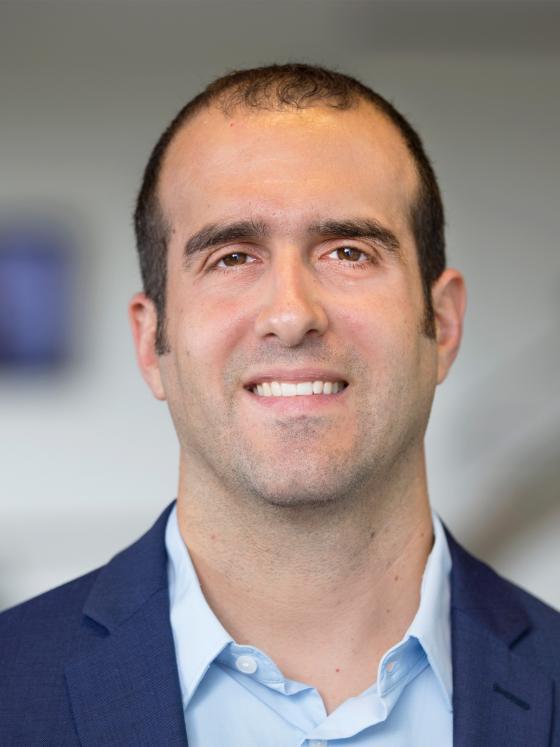
Dr. Mario Fabiilli is an Associate Professor of Radiology and Biomedical Engineering at the University of Michigan. His lab develops ultrasound-based therapies with a focus on biomaterials for drug delivery and tissue regeneration, including strategies for blood vessel and bone growth. The Fabiilli lab leverages ultrasound-induced effects to non-invasively and spatiotemporally modulate both biochemical and biophysical signals to direct cell behaviors.
Source
1. R. W. Wood, A. L. Loomis, Philosophical Magazine and Journal of Science 4, 417 (1927).
2. R. Puts et al., Eur Cell Mater 42, 281 (2021).
3. S. R. Burks et al., PloS one 6, e24730 (2011).
4. S. R. Burks, A. Ziadloo, S. J. Kim, B. A. Nguyen, J. A. Frank, Stem Cells 31, 2551 (2013).
5. M. Bez et al., Science translational medicine 9, (2017).
6. M. Bez et al., Molecular therapy : the journal of the American Society of Gene Therapy 26, 1746 (2018).
7. O. D. Kripfgans, J. B. Fowlkes, D. L. Miller, O. P. Eldevik, P. L. Carson, Ultrasound Med Biol 26, 1177 (2000).
8. H. Jin et al., Journal of Controlled Release 338, 773 (2021).
9. L. Huang et al., Acta Biomater 129, 73 (2021).
10. M. Aliabouzar et al., Ultrasonics sonochemistry 66, (2020).
11. E. Farrell et al., Acta Biomater 138, 133 (2022).
12. M. Aliabouzar et al., Acta Biomater 164, 195 (2023).
